| Multiresolution Characterization
of Interictal Epileptic Spikes based on a Wavelet Transformation
A. Barreto, N. Chin., J. Andrian J. Riley
Department of Electrical Engineering, Florida
International University
University Park, Miami, Florida, 33199
Abstract - A wavelet transformation is applied to electrocorticogram
(ECoG) records from epileptic patients. The temporal sharpness associated with interictal
spikes at different resolutions is observed and two ways for representing the
multiresolution sharpness of the spikes are proposed.
I. Introduction
In addition to the characteristic electrographic
bursts of abnormal activity that are recorded when epileptic patients experience a seizure
(ictal episode) , the electroencephalogram (EEG) of epileptics will normally display
isolated sharp transients or "spikes" in some locations of the brain. These
interictal spikes are a complementary source of information in the diagnosis and
localization of epilepsy.
In particular, when a prospective candidate for seizure surgery is
studied with long-term video/EEG monitoring, both the ictal (electrographic seizures) and
interictal (spikes) manifestations of epilepsy are scrutinized to determine the nature
and, in some cases, the localization of a focus of epilepsy. It is for this kind of
scenario that an automatic or semiautomatic method for interictal spike detection has been
sought for several decades.
Numerous attempts have been made to define a reliable spike detection
mechanism. However, all of them have faced the lack of a specific characterization of the
events to detect. One of the best known descriptions for an interictal "spike"
is offered by Chatrian et al. [1]: " transient, clearly distinguished from background
activity, with pointed peak at conventional paper speeds and a duration from 20 to 70
msec...". This description, however, is not specific enough to be implemented into a
detection algorithm that will isolate the spikes from all the other normal or artifactual
components of an EEG record. Some approaches have concentrated in measuring the
"sharpness" of the EEG signal, which can be expected to soar in the "pointy
peak" of a spike. Walter [2] attempted the detection of spikes through analog
computation of the second time derivative (sharpness) of the EEG signals. Smith [3]
attempted a similar form of detection on the digitized EEG signal. His method, however
required a minimum duration of the sharp transient to qualify it as a spike. Although
these methods involve the duration of the transient in a secondary way, they fundamentally
consider "sharpness" as a point property, dependent only on the very immediate
context of the time of analysis. More recently, an approach has been proposed in which the
temporal sharpness is measured in different "spans of observation", involving
different amounts of temporal context [4]. True spikes will have significant sharpness at
all of these different "spans". The promise shown by that approach has
encouraged us to use a wavelet transformation to evaluate the sharpness of EEG signals at
different levels of temporal resolution. We expect that, as in the previous study
mentioned above, the consistency of the sharpness displayed by the spikes across different
resolution levels will set them apart from other EEG transients. If this is the case a new
specification for interictal spikes, in terms of their characteristic multiresolution
sharpness, can be put forth.
II. Wavelets
When a signal is transformed into a representative
set of wavelet coefficients, each dilation represents a band-pass filtering of the input
signal corresponding to some specific scale which innately provides a useful mapping of
important signal features at different scales. This enables a more advanced analysis and
understanding of the signal through a more complete representation. Alternatively, this
can be seen as a type of template matching of important signal characteristics at
different scales (dilations) while maintaining the fundamental morphology of the wavelet.
There are many suitable wavelets which can be used such as those
developed by Mallet, Daubechies, and Morlet [5,6]. The particular wavelet function which
was used here, given below by Equation 2.1, is an offspring of Morlet’s wavelet.
 (2.1) (2.1)
In this case, the wavelet function y (t) is
admissible when a =s = , and b such that the function in (2.1) is zero. Allowing the parameter of
dilation, "a", to be inversely proportional to the harmonic of interest this
transformation can be accomplished through a discrete convolution of the time varying
signal with the wavelet function y *(t/a). Note that this
requires that c=2p so that the dilations of the wavelet be a
function of the frequency , and b such that the function in (2.1) is zero. Allowing the parameter of
dilation, "a", to be inversely proportional to the harmonic of interest this
transformation can be accomplished through a discrete convolution of the time varying
signal with the wavelet function y *(t/a). Note that this
requires that c=2p so that the dilations of the wavelet be a
function of the frequency  .
This is relevant since the parameter 'a' is just a scale for dilation so that establishing
this as the sweep frequency is a valid and necessary step [7,8]. Ultimately, the
multiresolution transformation generates an alternative representation for interpreting
spikes through the progression of morphological variability’s across many scales,
which distinguishes them from noise and background signals. .
This is relevant since the parameter 'a' is just a scale for dilation so that establishing
this as the sweep frequency is a valid and necessary step [7,8]. Ultimately, the
multiresolution transformation generates an alternative representation for interpreting
spikes through the progression of morphological variability’s across many scales,
which distinguishes them from noise and background signals.
For a function to be considered for use as a wavelet it is required
that the function be admissible. This requires that:
 (2.2) (2.2)
where y (w) is the Fourier
transformation of y (t), and Cg is the admissibility constant
[9]. This constant is required to be finite to allow for inversion of the wavelet
transformation. Any function which satisfies this constraint can be called a mother
wavelet and since Cg is finite then the mean value of the mother wavelet in time is zero
so that:
 (2.3)
(2.3)
To generate the wavelet transform,
W(b,a), of a signal, s(t), requires that the analyzing wavelet be convolved with the
signal as given in Equation 2.4 below.
 (2.4) (2.4)
Here, ‘b’ the parameter of translation is responsible for
localization in time and 'a' the parameter of dilation is responsible for localization in
frequency. This is accomplished discretely by sampling the input with a period T at least
two times larger than the highest harmonic of interest in s(t) such that:
 (2.5) (2.5)
Finally, this can be rewritten as:
 (2.6) (2.6)
Thus, the wavelet must be convolved with the input signal by adjusting
the parameter of translation 'b' and adjusting the sweep frequency for each iteration
(scale). For this particular application the most suitable wavelet function has a shape
which resembles the fundamental morphology of an interictal spike.
III. Application of Wavelets to Epileptic Spike Detection
The first step in applying the wavelet
transformation of Equation 2.6 to the detection of epileptic spikes was in defining the
most suitable wavelet parameters. Initially, with b =0 in
Equation 2.1, a pseudo wavelet was constructed and tested for sensitivity to spikes across
many scales. These tests were performed on portions of signals recorded from the brain of
epileptic patients with implanted electrodes, such as the one shown in Figure 3.1.

Figure 3.1 : ECoG Segment with Spike
It was determined that the wavelet transformation could be adjusted for
sensivity to change through the damping parameter, s , and for
localization in frequency through the harmonic analyzing parameter c. Thus, for each
iteration of the wavelet transformation the damping function and the specific frequency of
concern were varied.
The initial simulations involved varying different values of s and c. The results which are shown below in Figures 3.2 & 3
were examined and it was established, subjectively, that the optimal values where c=6 and s =3.5. As evidenced in the central traces of Figures 4.2 & 3
these values produced the most discernible output for the spike from Figure 3.1.

Figure 3.2 - Varying  parameter parameter

Figure 3.3 - Varying c parameter
The use of the pseudo wavelet in this preliminary stage is justified by
its morphological similarity with the true admissible wavelet, as shown in Figure 3.4.

Figure 3.4 - Pseudo and Admissible Wavelets
In addition, the values found for the parameters
made the pseudo wavelet closely approximate the admissible wavelet. Therefore, the rest of
the study proceeded with the use of the admissible wavelet. oNEC
IV. Results
Applying a wavelet decomposition that involved ten
different frequencies (1/a), (from 1 Khz to 10 Khz), a two dimensional output was obtained
from each ECoG segment used as input. The x axis of this output represented the sample
number, i.e. time. The y axis of the two dimensional output was associated with the
different wavelets from the set that was applied to the ECoG data. So, the two dimensional
output offered a representation of the ECoG signal similar to the "spectrogram"
used in the analysis of speech signals, except for that the decomposition is not based on
sinusoidal components, but it refers to the ten wavelet components used. Observation of
these two-dimensional outputs confirmed that epileptic spikes would have high outputs for
a larger number of wavelets in the set. On the other hand, background activity would
normally display high outputs for the lower frequency wavelets (wide wavelets) and
transients of artifactual origin would not have high output for many of the resolution
levels employed. Since the narrower wavelets would inherently yield a significantly lower
output than the wider ones, the two dimensional output was normalized so that the highest
value resulting from each wavelet dilation would be made 1.Figure 4.1 shows a 2-second
ECoG segment with a clear interictal spike. Figure 4.2 displays its normalized wavelet
decomposition.
Figure 4.3 shows another segment of ECoG data with some spikes and
other transients. Figure 4.4 shows the corresponding two-dimensional output of the wavelet
transform.

Figure 4.1 - ECoG segment
with spike

Figure 4.2 - Wavelet transformation of Figure 4.1
In it, the spikes show consistent large outputs throughout the wavelet
set, i.e. they have sharpness at several different resolutions. On the other hand, the
transients in the second half of the segment only yield a large output for some subsets of
the wavelets, i.e., they only have sharpness at certain resolutions. To summarize these
differences and enable a detection mechanism we first suggest the point-to-point
multiplication of the results for several wavelet dilations, so that only the features
that are sharp at all of those resolution levels will be represented by large product.
Figure 4.5 illustrates this option for the isolation of interictal events.
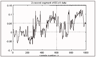
Figure 4.3 - Segment with spikes and other
transients

Figure 4.4 Wavelet transformation of Figure 4.3

Figure 4.5 - Product of the wavelet coefficients for 3, 5 and 7
Khz(solid) and 3 and 5 Khz only(dashed)
Another possibility that we propose is to use the outputs obtained at
three different resolution levels as the x, y and z coordinates in a parametric plot. In
this way, only features that have sharpness at all three of those resolution levels will
result in large orbits, away from the origin of the coordinate system. Initially, a
spherical boundary can be set around the origin to act as a threshold for features that
may be interictal events. This form of display is particularly interesting, since not only
the farthest position reached by an orbit, but also the specific trajectory could be used
to classify features. Figure 4.6 shows one such parametric plot, for the data in figure
4.3.

Figure 4.6 Parametric plot of the wavelet
coefficients at 3, 5 and 7 KHz
V. Conclusions
Through this study we have found that a wavelet
transformation is capable of separating a time series, such as the ECoG from an epileptic
patient, according to the sharpness of the signal at different temporal resolutions. We
have also observed that interictal spikes display significant sharpness at several
resolution levels, while other artifactual transients and background features normally do
not show consistent sharpness at the resolution levels chosen. This new characterization
of the spike in the frame of multiresolution analysis may be used to develop a detection
signal derived from the output of the wavelet transformation as a product of the outputs
at several resolution levels or using these as coordinates for a parametric plot.
VI. References.
[1] G. Chatrian et al., "A glossary of terms
most commonly used by clinical electroencephalographers", Electroenceph. and Clin.
Neurophysiol., 1974, 37:538-548..
[2] D. Walter et al., "Semiautomatic quantification of sharpness
of EEG phenomena". IEEE Trans. on Biomedical Engineering, 1973, Vol. BME-20, pp.
53-54.
[3] J. Smith, "Automatic Analysis and detection of EEG
Spikes", IEEE Trans. on Biomedical Engineering, 1974, Vol. BME-21, pp. 1-7.
[4] A. Barreto et al., "Intraoperative Focus Localization System
based Spatio-Temporal ECoG Analysis", Proc. XV Annual Intl. Conf. of the IEEE
Engineering in Medicine and Biology Society, October, 1993.
[5] Daubechies, Ingred. Ten Lectures on Wavelets SIAM (Society
for Industrial and Applied Mathematics), Philadelphia, Pennsylvania, 1992.
[6] S. Mallat, "A Theory for Multiresolution Signal Decomposition:
The Wavelet Representation", IEEE Trans. on Pattern Analysis and Machine
Intelligence, vol. 14, pp710-732, July 1992.
[7] Mireille Barrat and Olivier Lepetit, "Calcul Rapide de la
Transformee en Ondelettes; Fast Processing of the Wavelet Transform" Traitement du
Signal, volume 8, No 1, Feb 1990.
[8] Oinis Chaari and Michel Meunier, "A Recursive Wavelet
Transform Analysis of Earth Fault Currents in Petersen Coil Protected Power Distribution
Networks" , 1994, IEEE. Wavelets
[9] Lora G. Weiss, "Wavelets and Wideband Correlation
Processing", I.E.E.E. Signal Processing Magazine, Vol.11 No.1 1994.
|